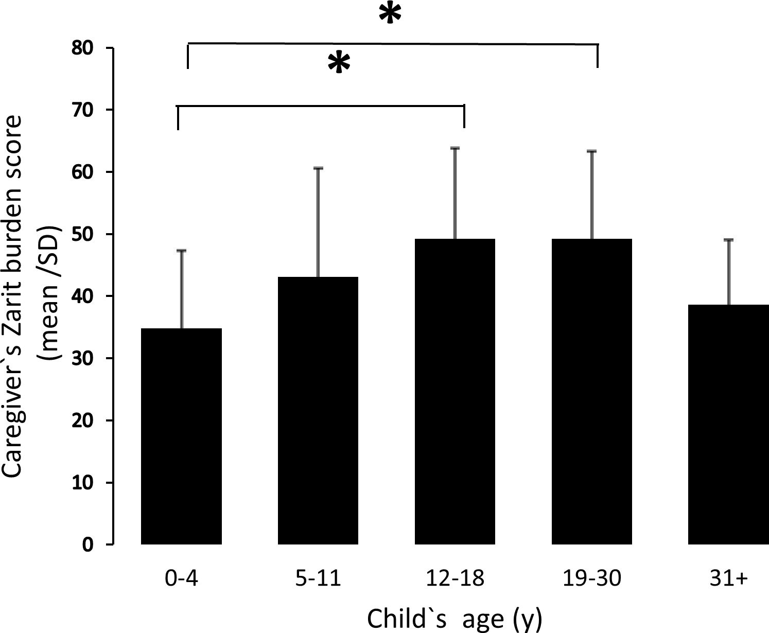
Accelerated ageing in PWS
NOV 2019 – With better management, people with PWS are living longer today, but the number of people known to our Association aged over 50 years is still relatively small. There is hope that this will change in the future with the advantages of early diagnosis and growth hormone treatment, but it has long been hypothesised that premature ageing occurs in PWS.
It has been observed that physical appearance can change earlier (mid to late 30’s) and that problems associated with older age in the general population can appear earlier. It is thought that there might be lots of health and wellbeing factors influencing to what extent the ageing process is accelerated, but special health checks for adults with PWS from the age of 40 years have been advised since the publication of a Dutch study by Sinnema M. et al., 2012. This study highlighted a need for regular checks which screen in particular for cardiovascular disease, diabetes, dermatological problems, orthopaedic problems including osteoporosis, sleep and psychological functioning. It was suggested that surveillance from 40 years onwards would ensure that intervention and support is offered at the earliest possible time.
Increased brain-age in young adults with PWS
To date, exact causes of accelerated ageing remain unconfirmed. There are competing hypotheses (arrested development vs. premature aging) which aim to explain structural brain abnormalities in PWS. Earlier this year, a study led by researchers at Imperial College London and the University of Cambridge (Azor AM, Cole JH, Holland AJ, et al., 2019) indicated that PWS is associated with an increased brain-age from early adulthood. They found that individuals with PWS had a brain age that more closely resembled healthy adults on average 8.74 years older than their chronological age and concluded that participants with PWS showed signs of premature brain aging and early onset atrophy, rather than a fundamental arrest of brain development. They stated that patterns of brain structure more closely resembled healthy older brains rather than healthy younger brains, which might be indicative of premature neuronal loss and atrophy.
Structural brain abnormalities and abnormal development in certain brain areas are widely reported and this study suggests that there may be neurological implications of the syndrome in older age. Few of the previously reported structural brain abnormalities were detected in the participant group for this study and so they were able to distinguish such structural abnormalities from premature brain aging. More localised examination of structural brain differences revealed other differences. Interestingly, the higher brain age in PWS was not related to changes in grey matter and white matter volumes which did not differ between controls and PWS groups.
They also found that premature brain ageing was not associated with current BMI, use of hormonal or psychotropic medications, gender or IQ. Further studies were deemed necessary to clarify the relationship with obesity, age of onset and duration. It should be noted that the vast majority of subjects in this study had a paternal chromosome deletion, with only 1 out of 20 having mUPD.
Evidence for accelerated biological ageing in young adaults with PWS
In November this year, another study by Donze, Stephany H et al. (2019) measured leukocyte telomere length (LTL) 1 from blood samples (a marker of biological age) in 47 young adults with PWS and compared LTL to healthy young adults of similar age. They found that young adults with PWS have significantly shorter median LTL compared to age-matched healthy young adults and GH-treated healthy young adults born short for gestational age, which provided evidence for accelerated biological ageing in PWS. Besides the finding that LTL is shorter in young adults with PWS, they found a tendency toward an association between a shorter LTL and a lower total IQ, which might imply a role of LTL in cognitive functioning in PWS. They found no significant difference in median LTL between males and females, possibly due to generally low levels of oestrogen in females with PWS, which would usually lengthen LTL. There were also no differences observed between genetic subtypes of PWS, but this was unexpected because a previous study by Whittington et al 2 reported on 26 adults with PWS over the age of 40 years and observed that when significant deterioration in executive functioning occurred with possible dementia, all cases had UPD. Psychiatric disorders are also reported more commonly in people with UPD and a higher level of psychosocial stress is associated with shorter telomeres. The researchers explained that it is difficult to compare the 2 studies because the group studied by Whittington et al were considerably older and none were treated with GH.
All the young adults who participated in this study were treated with long-term GHT. There was no association between LTL and the duration or dose of GH in young adults with PWS, which is reassuring in regard to speculation of negative effects of GH on LTL. The researchers suggest it is possible that GH treatment might positively influence LTL by improving body composition. Early diagnosis, intervention and GH treatment has brought about a new generation of young adults with PWS without severe obesity for whom it is suggested may have had shorter LTL had they not been treated with GH as they would have had a higher BMI and FM%. In this Dutch study they were not able to compare LTL in GH-treated young adults with PWS to untreated young adults with PWS so more research is needed on LTL across the life course to be able to determine its role in ageing for both GH-treated and untreated people with PWS.
In conclusion, the researchers discuss that even though none of the genes in the PWS region on chromosome 15 are associated with telomere homeostasis, several clinical features of people with PWS are associated with an increased risk of shorter telomeres, including obesity, reduced physical activity, and increased psychosocial stress. Furthermore, adults with PWS are prone to develop T2 diabetes and cardiovascular disease in early life and several studies have shown that shortened telomeres are associated with an increased risk of these conditions. The study concluded that shorter telomeres might play a role in the premature aging in PWS, independent of GH, and suggested studies are needed to further determine the influence of LTL on aging, cognitive functioning and brain development in PWS.
- Telomeres are short DNA-protein structures forming a protective cap at the ends of each chromosome. They play a role in protecting our genome from degradation and loss of essential DNA. As people grow older, their telomeres shorten because a small portion of telomeric DNA is lost during the normal process of cell division to replicate chromosome DNA. Eventually telomeres become too short to do their job, causing cells to age and stop functioning properly. Lifestyle factors, such as smoking, obesity, stress or lack of sleep, can accelerate telomere shortening, whereas healthy diet, exercise, and stress reduction can lengthen them.
- Whittington, J E et al. “Ageing in people with Prader-Willi syndrome: mortality in the UK population cohort and morbidity in an older sample of adults.” (2015)